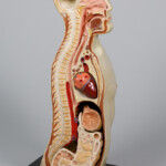
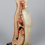
Photos: WUM Photomedical Department
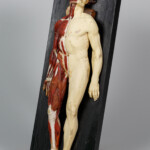
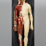
We have just received new deposits – these are pre-war anatomical models that were lent to us thanks to the courtesy of the head of the Department of Human and Clinical Anatomy, prof. Bogdan Ciszek. Made of plaster (often with the addition of “mache” paper) and hand-painted, such models became popular teaching materials for the study of anatomy in the early 20th century and a practical replacement for hard-to-reach human corpses.
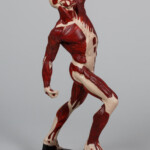
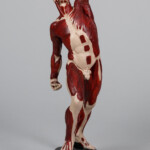
All three models will be a great addition to our permanent exhibition, which opens in winter 2021. One of them is the male figure shown in photos 1 and 2 with exposed muscles. The left arm is raised with the arm bent at the elbow. The right arm is slightly bent, pointing down with the palm of the hand up. The leg position causes the hips to twist. This position allows one to define and visualize detailed muscle groups of the arms and legs, upper chest and hips both when stretched and tense (on the left side of the figure), and flexed and slightly relaxed (on the right side of the model). From the 18th century onwards, this kind of representation of an anatomically refined “muscle man” has been reffered to by the French term écorché (skinned) in both art history and medicine.
The earliest confirmed artistic representations of écorchés come from the Renaissance period. Many écorché drawings by Leonardo da Vinci (1452–1519) have survived, for example the study of the arm with its arm stretched downwards in four positions (1509–1510. Other famous examples are Jean Antoine Houdon’s Ecorche from 1767, St. Bartholomew “Marco d ‘Agrate from 1562 or” Smugglerius “by William Pink from 1864.
All these famous Ecorches along with photos have been posted in the Art — Works of Art section of this website.


