
Galen (also known as Galenus) was a Greek anatomist, writer and philosopher who was considered the most famous physician in the Roman Empire; his theories dominated European medicine for almost 1,500 years. He was born circa 129 CE in Pergamum (modern-day Turkey), and studied in Greece, Alexandria and Asia Minor. After completing his studies, he became chief physician of the gladiators’ school in Pergamum, where he gained a great deal of experience in the treating of wounds. At the beginning of 160 CE, Galen moved to Rome where he spent the remainder of his life. He became physician to the Emperors Marcus Aurelius, Commodus and Septimus Severus. Galen was the originator of the experimental method in medical research and throughout his life dissected animals in his quest to understand how the body functions. Some of his anatomical and physiological observations were accurate – for example, he demonstrated the muscles and nerves which control the vocal chords and breathing; he proved that spinal cord injuries caused loss of movement and feeling; that urine was formed in the kidney (and not in the bladder as was commonly believed); he also identified 7 of the 12 cranial nerves – such as the optic nerve and vestibulocochlear nerve (which transmits sound and balance (equilibrium) information from the inner ear to the brain). Nevertheless his research was limited by the fact that in the times in which he lived he was unable to perform autopsies on human bodies. Galen was a prolific author, with hundreds of medical treatises to his name, many of which have unfortunately not survived to this day. In his work he compiled all significant Greek and Roman medical thought to date, and added his own discoveries and theories. His achievements reigned supreme for fifteen centuries after his death in 216 CE. Many of his theories were not disproved until the Renaissance.

Mondino de Luzzi (also known as Mundinus) was born in Florence in 1270 and studied medicine at the University of Bologna, with which he was associated for the whole of his life. He conducted his first public dissection in Bologna in 1315 – the first recorded dissection of a human body in more than 1,700 years. His publication Anathomia corporis humani, written in circa 1316, was the first book in Europe devoted solely to anatomy that had been written since antiquity. The information it contained was based on the dissection of a human body and took the form of a textbook which – step by step – showed the various stages of dissecting a body, starting from the abdomen (which contained organs that were the first to decompose) up to the head. For 250 years it was the dominant source of knowledge on anatomy until the Flemish anatomist Andreas Vesalius published his renowned work De Humani Corporis Fabrica. Much of the content in Mundius’ book comes from the teachings of Galen, Aristotle and Hippocrates, and therefore, to some extent, it repeated errors that had been made in the past, and which were criticized by Vesalius 200 years later. Anathomia, among other things, includes descriptions of surgical procedures for hernias and cataracts as well as descriptions of cranial sutures and the brain. Despite its imperfections, Mundinus’ publication was the most frequently used anatomical text for the next 250 years.

Leonardo Da Vinci became interested in anatomy through painting. The Renaissance celebrated the beauty of the human body and Da Vinci wanted to understand its structure, proportions and depict it as realistically as possible in his paintings. From 1489 he began anatomical studies,focusing on the human skull – the profiles, with the various cross sections are carefully drawn and shaded, and the correct alignment of the nerves and blood vessels inside it is shown. In 1506 he performed his first dissection of the human body outlining the various stages of the process. He described atherosclerosis for the first time and was the first person in the history of medicine to describe cirrhosis of the liver. He was allowed to examine more than 30 cadavers at the University of Padua. In the years 1510–1511 he prepared a series of sketches, most of which were double-sided, of nearly 240 drawings, which was known as Anatomical Manuscript A. Da Vinci focused on an analysis of the bones and muscles, detailing each bone of the human body and individual groups of muscles. He sketched the first known representation of the human spine, with its typical curvature and the correct number of vertebrae. He also described the appendix – in his day the inflammation was treated with castor oil; the removal of an inflamed appendix was recorded for the first time by the London doctor Claudius Amyand in 1735. Because he was not a doctor, the Pope refused to give him further access to cadavers so he had to continue his anatomical research on animal organs. He therefore used bulls’ hearts to study the circulatory system. Contrary to Galen’s traditional idea, he recognized that the human heart was a muscle with four chambers (the systemic circulation of the blood was first described in detail in 1628 by the English physician William Harvey). In order to make his famous drawing of an embryo in the womb, Da Vinci dissected a pregnant cow and based on the observations he made his famous drawing of an embryo in its mother’s womb. Da Vinci’s discoveries could have revolutionized anatomy and medicine in general, but unfortunately his notes were not published in his lifetime. Many of his early findings were not described again until 250 years later. After his death in 1519 many of his drawings were lost. The remainder were not rediscovered until the 20th century and are now largely part of the collection of the British Royal Family.

Alessandro Achillini was born in 1463 in Bologna and during his lifetime was known mainly as a philosopher. Today, however, he is remembered for his considerable activities in research on the human anatomy. In 1484 he obtained a doctorate in philosophy and medicine from the University of Bologna and Bologna was where he spent the greater part of his life. His two best known works are De humani corporis anatomia and Annotationes anatomicae. In his work he gave a detailed description of the veins in the arm, the fornix of the brain, the so-called infundibulum and trochlear nerve. He also accurately described the ileocecal valve which was later described by Costanzo Varolio and Gaspard Bauhin. Finally, Achillini is credited with the first description of two of the three bones of the inner ear – the malleus and incus. He is widely considered to be the greatest researcher on the human brain in the 15th century. Achillini was the first person to demonstrate a few of Galen’s mistakes, including the seven bones comprising the tarsus (the middle part of the foot). Galen claimed there were nine. Among the Italian anatomists from before the era of Vesalius (Alessandro Benedetti, Gabriele Zerbi, Berengario da Carpi and Niccolo Massa) the influence of Achillini’s work was not great mainly because his work was not illustrated and the mediaeval terminology he used was derived mainly from the texts of Mondino de Luzzi and was not transparent and intermixed with Arabic terminology. He died in 1512.

Bartolomeo Eustachi was born in 1500. He was one of the founders of the science of human anatomy and one of the most important scientists who contributed to the development of Renaissance medicine. He was also a 16th century contemporary of Vesalius, although the two anatomists did not like one another (Eustachi was an adherent of Galen’s teachings). He spent most of his working life in Rome, where he taught anatomy and performed dissections of cadavers in hospitals. In 1562 and 1563 Eustachi published a unique series of treatises on the kidneys (De renum structura), the inner ear (De auditus organis) and the venous system (De vena quae azygos graecis dicitur). They were published in Opuscula anatomica (1564). The treatise on the kidneys was the first work to be devoted to that organ. His treatise on the inner ear contained an accurate description of the tube that now bears his name – the Eustachian tube. Eustachi was the first person to study in detail the anatomy of the teeth in his treatise Libellus De Dentibus. In 1552 Eustachi prepared a series of 47 anatomical drawings depicting human skeletons and muscles of which only eight were published in his lifetime. Eventually all the plates ended up in the Vatican Library. In the 18th century the pope’s physician, Giovanni Maria Lancisi, added explanations to some of the earlier unpublished plates and published the complete set in 1714 (140 years after the author’s death) under the title Tabulae anatomicae Bartholomaei Eustachii. Although Eustachi’s drawings were not as fine from an artistic perspective as those prepared by Vesalius in his De Humanis Corporis Fabrica, Eustachi is sometimes far more accurate in his descriptions. Had the complete set of plates been published ten rather than 140 years after Vesalius’ publication, it is very likely that both of them would have been regarded as the co-founders of modern anatomy. Eustachi died on 3 June 1657.

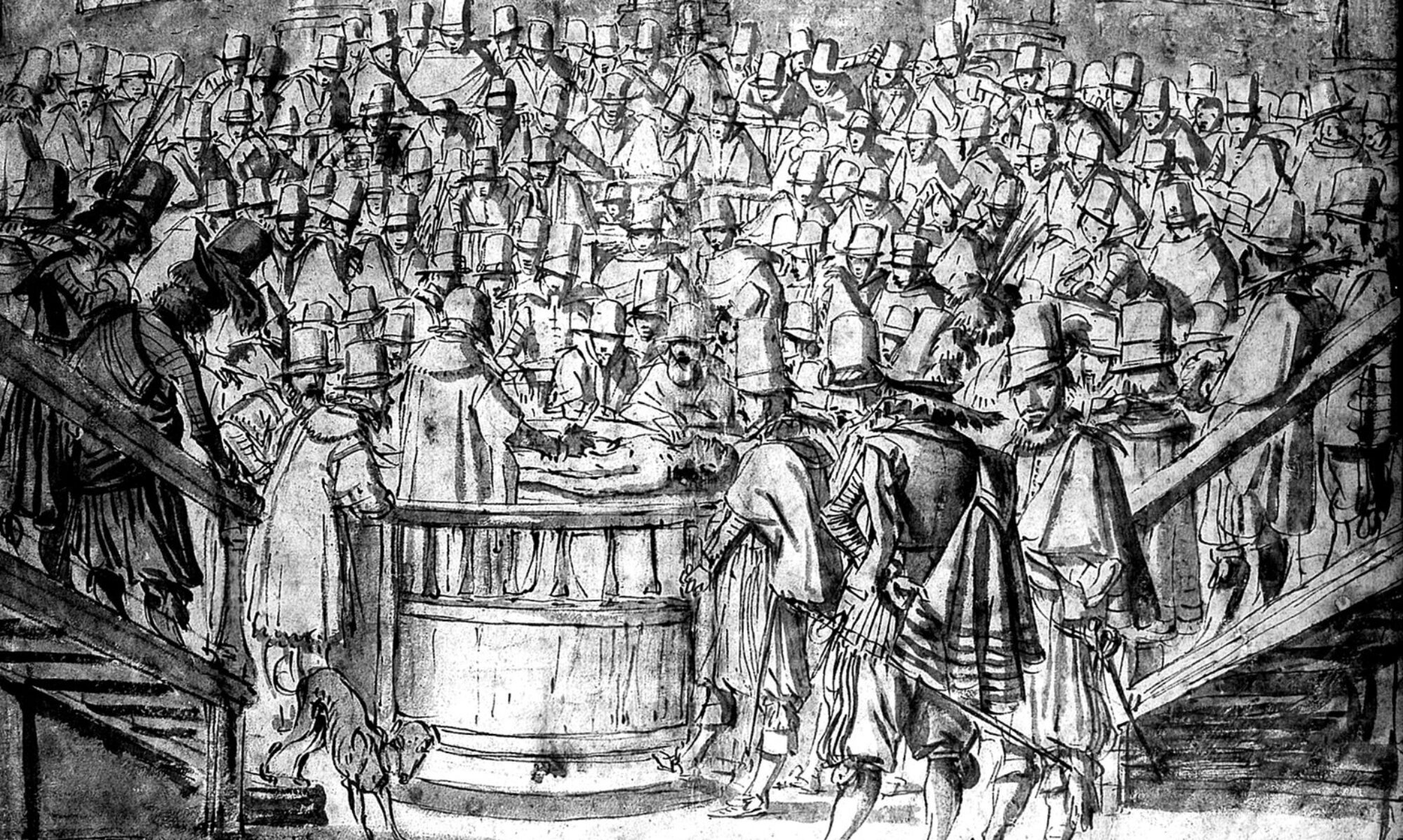
Andreas Vesalius was born on 31 December 1514 in Brussels in the Duchy of Brabant which was then part of the Holy Roman Empire (later Belgium). He studied medicine in Paris but was forced to leave before having completed his studies due the Holy Roman Empire declaring war on France. He then studied at the University of Leuven before transferring to Padua where he began his medical degree. After graduating in 1537 he took up the position of professor of surgery and anatomy in Padua. Thanks to having constant access to the corpses of convicts, Vesalius was able to perform multiple and comparative dissections on human cadavers. This was in stark contrast to Galen, who had been the main authority on anatomy to date and who, for religious reasons, confined himself to dissecting the corpses of animals, mainly monkeys. Vesalius was aware that Galen’s observations and his own differed and that the human anatomy differed from that of apes. In 1543 Vesalius published his famous De Humani Corporis Fabrica. It was richly illustrated (most probably by Steven van Calcar, a pupil of Titian) which transformed anatomy into a subject that was based on empirical observations taken directly from the dissection of human bodies. In recognition of his groundbreaking work, Vesalius was invited to become imperial physician at the court of Emperor Charles V, and in 1555 he continued working for Charles’ son, Phillip II of Spain. In 1564 he went to the Holy Land and died on 15 October 1564 on the Greek island of Zakynthos while travelling back home.

William Harvey was an English physician who was the first to accurately describe the circulatory system, and he recognized that the heart was the organ which pumped the blood through the body. William Harvey was born in 1578 in Folkestone in Kent. He was educated at King’s College in Canterbury, then at the University of Cambridge and later studied medicine at the University of Pauda in Italy, where he became a student of the scholar and surgeon Hieronymus Fabricius. Fabricius, fascinated by anatomy, recognized that the human body has non-return valves, but did not know how they functioned. This basic work by Fabricius provided Harvey with the basis to solve the puzzle of the role of the valves in the circulation of blood in the body. After returning from Italy in 1602, Harvey was appointed physician at St Bartholomew’s Hospital in London in 1609. In 1618 he was appointed physician to King James I and continued as physician to his son Charles upon his accession to the throne. Harvey continued performing dissections on animals. In 1628 he published his theories in a book entitled Exercitatio Anatomica de Motu Cordis et Sanguinis in Animalibus (Anatomical Exercise on the Motion of the Heart and Blood in Animals), in which he gave a scientific explanation of the circulation of the blood. This put an end to the influence of Galen’s teachings on anatomy. Harvey was the first to suggest that mammals reproduced by fertilizing an egg with a sperm. Another two hundred years elapsed before an egg was finally observed in a mammal, but Harvey’s theory did gain credibility during his lifetime. He died on 3 June 1657.