
We are pleased to announce that our first part of permanent exhibition will be opened to visitors on May 18. The exhibition will be available to you on Mondays – Fridays from 9.30 a.m. to 3.30 p.m.
We look forward to see you!
MUZEUM HISTORII MEDYCYNY WUM
We are pleased to announce that our first part of permanent exhibition will be opened to visitors on May 18. The exhibition will be available to you on Mondays – Fridays from 9.30 a.m. to 3.30 p.m.
We look forward to see you!
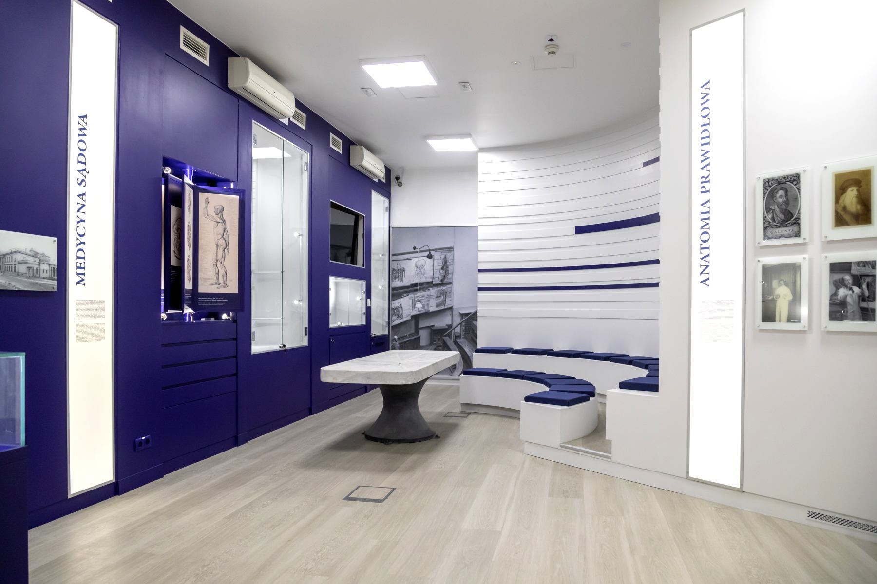
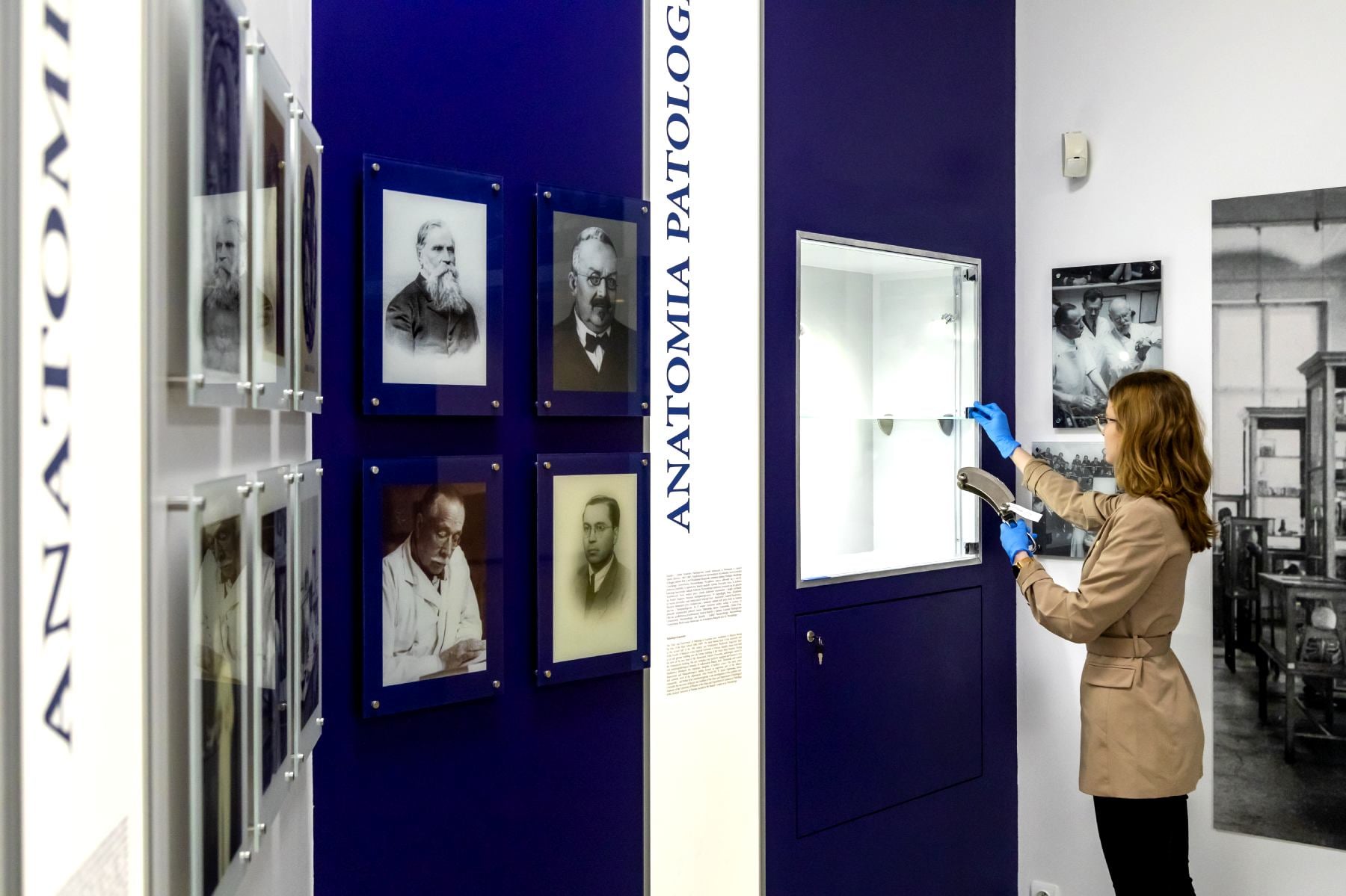
Today we are delighted to share with you the exciting news about a very important event in the history of our Museum. The completion of the first showroom of our anatomical theater has just finished, and thus we are starting the arrangement of our new exhibition. The display is being prepared, and more artefacts are taking their rightful place in museum display cases. Another task is to perform 3D mapping – after its completion and after lifting the restrictions, we will be able to welcome you at the first permanent exhibition of our museum. The ceremonial opening of the new space will take place before Easter – the report from this event will be available through all communication channels of our museum.

 As part of our project ” Uncover the secrets of the human body in an anatomical theatre of the 21st century” we have prepared an educational pack for you, thanks to which you will be able to train your brain and consolidate the knowledge about anatomy acquired through our lectures, workshops and website. Already printed educational brochures are safely stowed in our museum, waiting patiently for the university gates to be opened to the public. For now, we provide you with a digitalized version. We wish you a lot of fun!
The folder in pdf format is available for download HERE:
Prepared by: Diana Święcka
Graphics and typesetting: Piotr Berezowski
The project “Uncover the secrets of the human body in an anatomical theater of the 21st century” is co-financed by the “Social Responsibility of Science” programme of the Ministry of Science and Higher Education
We are very pleased to announce the implementation of the next stage of our project “Learn the secrets of the human body in the anatomical theater of the 21st century”. As part of the ministerial subsidy, a set of educational models was purchased, which will be widely used in promoting knowledge about the structure of the human body. Our new acquisitions will be available to both students preparing for academic classes and all visitors to the newly established anatomical museum theater. Phantoms will be used during workshops and educational lectures aimed at children, adolescents and adults as well. We are therefore looking forward to the opening – as soon as the epidemiological situation allows – our new educational space, and we encourage all interested students to apply to the Museum of the History of Medicine of the Medical University of Warsaw to be able to take advantage of our new acquisitions (muzeum@wum.edu.pl) ).
We cordially invite you to see our gallery showing all the phantoms!
The anatomical models were purchased as part of the project ” Uncover the secrets of the human body in an anatomical theater of the 21st century “, co-financed by the program “Social Responsibility of Science” run by the Minister of Science and Higher Education.


We are pleased to present you “Milestones in the history of anatomy” – animation that was created as part of the project “Uncover the secrets of the human body in an anatomical theater of the 21st century”. This looped, short movie will be presented in our newly created museum educational space.
The film is full of drastic scenes, dramatic plot twists, but above all, delivers a solid dose of knowledge about the history of human anatomy.


Photos: WUM Photomedical Department
We have just received new deposits – these are pre-war anatomical models that were lent to us thanks to the courtesy of the head of the Department of Human and Clinical Anatomy, prof. Bogdan Ciszek. Made of plaster (often with the addition of “mache” paper) and hand-painted, such models became popular teaching materials for the study of anatomy in the early 20th century and a practical replacement for hard-to-reach human corpses.
All three models will be a great addition to our permanent exhibition, which opens in winter 2021. One of them is the male figure shown in photos 1 and 2 with exposed muscles. The left arm is raised with the arm bent at the elbow. The right arm is slightly bent, pointing down with the palm of the hand up. The leg position causes the hips to twist. This position allows one to define and visualize detailed muscle groups of the arms and legs, upper chest and hips both when stretched and tense (on the left side of the figure), and flexed and slightly relaxed (on the right side of the model). From the 18th century onwards, this kind of representation of an anatomically refined “muscle man” has been reffered to by the French term écorché (skinned) in both art history and medicine.
The earliest confirmed artistic representations of écorchés come from the Renaissance period. Many écorché drawings by Leonardo da Vinci (1452–1519) have survived, for example the study of the arm with its arm stretched downwards in four positions (1509–1510. Other famous examples are Jean Antoine Houdon’s Ecorche from 1767, St. Bartholomew “Marco d ‘Agrate from 1562 or” Smugglerius “by William Pink from 1864.
All these famous Ecorches along with photos have been posted in the Art — Works of Art section of this website.


Photos: Wellcome Collection
One of the most famous eighteenth-century anatomical atlases, which was created with the participation of the graphic artist and anatomist Jacques Fabien Gautier d’Agoty (1716-1785). The main task of this work was to facilitate the study of anatomy for students of medicine, surgery, painting and sculpture. In the creation of this work and other anatomical atlases, Gautier d’Agoty collaborated with Jacques Francois Duverney, a Parisian surgeon and anatomy demonstrator at the Jardin du Roy. As far as the medical value of the content of the atlas is concerned, it was small in the eyes of specialists and has added nothing significant to science since the time of Vesalius. On the other hand, the illustrations that complemented it were extremely valuable. They were made using the mezzotint method, which Gautier d’Agoty learned from his master Jacob Christoph Le Blon, enriching them later with the addition of black. The anatomical pictures from Myologie complette aroused much controversy in the 18th century. They were attributed to sublime drama, artistic arrogance, and sometimes even an insult to Christian morality. The realistic close-ups of female and male genitals depicted next to each other were particularly controversial. In some cases, they were read as disguised personal erotic fantasies of the artist. It seems, however, that in this way he rather wanted to draw the attention of influential personalities to his works, which could have positively influenced the expansion of his printing activities. Today, due to the characteristic style and extremely original compositions, Gautier d’Agoty is considered to be the progenitor of surrealism.

On behalf of the Museum of the History of Medicine of the Medical University of Warsaw and the honorable speaker – dr hab. n. med. Tomasz Dzieciatkowski, we are honored to cordially invite you to an open lecture entitled “Viruses and epidemics – a trending topic”. It will be the first in a series of ten lectures carried out as part of the project “Learn the secrets of the human body in the anatomical theater of the 21st century”. Continue reading “Open lecture: “Viruses and epidemics – a trending topic””

The most famous anatomical textbook of the modern era. Written by Andreas Vesalius from Brussels, educated in Paris and professor in University of Padua. It summarizes the years of entire human research that led to the challenging of medical science in Galen’s day. It became possible thanks to the regular dissections of human corpses by Vesalius. The work of De Humanis Corporis Fabrica contains 273 illustrations. Their author was most likely Titian’s student Jan Stefan von Calcar. The characters presented by him are characterized by dynamics and, above all, diversity. Both the skeletons and flayed ecorché were captured in a pensive or dancing pose. All the drawings are distinguished by an extremely perfect observation of the muscular system, a precise capture of the structure of nerves, vessels and bones. No one before Vesalius has combined medicine and art so closely in one work, therefore the question is often asked who played a greater role in its creation, the doctor or the artist. The work, thanks to the print, was very quickly disseminated in Europe. It was enthusiastically received in many circles, but there were also voices of criticism that did not approve of the progress of science captured in it.

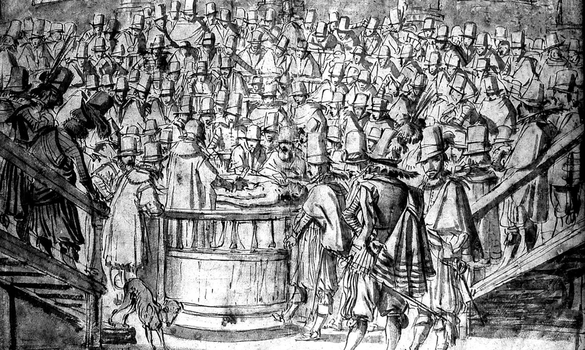
The work of Queen Jeanne de Bourgogne’s personal physician Guido de Vigevano (c. 1280 – c. 1349) was dedicated to the King of France, Philip VI. It is one of the most interesting 14th-century manuscripts dedicated to human anatomy. He refers directly to the Galen manuscripts and the teachings of the mentor Vigevano Mondino de Luzzi. Especially valuable are the illustrations contained in it, which were created thanks to the experience of this physician in carrying out an autopsy on human corpses, although they were officially prohibited during his lifetime. In one of the drawings the human body is even shown on the “autopsy” table, so that the corpse and the anatomist standing next to it occupy two planes next to each other. The next pictures show the anatomy of the abdomen, chest and head, and this is the most famous anatomy of a woman’s body with a visible “seven-chambered” uterus in reference to Galen’s hypothesis. In Vivegano’s work there are also drawings showing treatments carried out on living patients. All pictures are highly schematic and with low precision. In 1926, Vigevano’s work was reissued in color by the French librarian and historian of medicine Ernest Wickersheime.